How can you tell if you are hearing or seeing propaganda instead of reasoned debate or analysis?
If there is no mention of the child at all, not even a mention of a “fetus” or “embryo,” or if there is no photo of the unborn child, or even a written description of her appearance, that is propaganda.
If the unborn child is called a “fetus” or “embryo” that might be propaganda. “Fetus” is a proper medical term for an unborn child from 13 weeks gestation (11 weeks after fertilization). “Fetus” means “offspring” or “progeny.” The feminine is “parva fetus.” If abortionists want to use Latin to dehumanize the child, then the equivalent correct medical term for the mother would be “gravida.” There is no masculine equivalent, despite the wild imaginations of those who refer to “pregnant people” rather than “pregnant women.”
To imply that an unborn child is not a “child” but a “fetus” or an “embryo” is logically analogous to saying that “she is not a child, she is a toddler.” “Fetus,” like “toddler,” designates a specific phase of the state of “childhood.”
Photos of the unborn child at 11, 13, 17 and 25 weeks gestation are seen below.
I deposed an abortionist under oath. In addition to the 65,000 abortions he performed in Jacksonville, he also had a side business of fertility treatments. He published a huge ad in the Raleigh News & Observer for his fertility business. It was entitled “Mr. & Mrs. Johnson Meet Their Son For the First Time.” It included an ultrasound photo of a preborn child only six weeks after fertilization. I asked him how he could refer to him as a “son” when he aborted thousands of them at the same age. He answered that his staff wrote that ad. He didn’t like it!!
The writer, Paul Stam, served 16 years in the NC House, the last 10 as Republican Leader and Speaker Pro Tem. See Articles on www.paulstam.info for further information. He can be reached at paulstam@stamlawfirm.com.
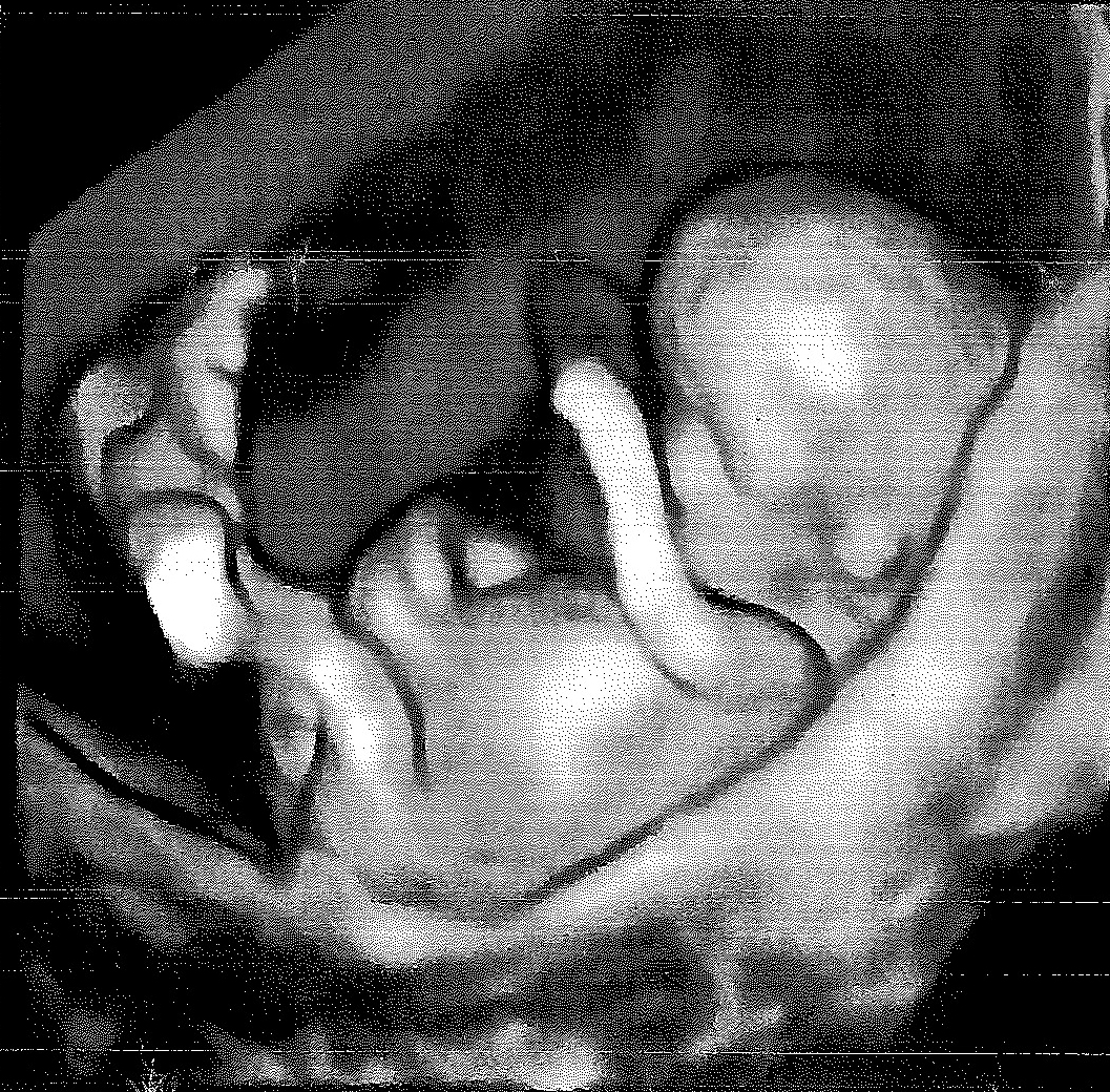
Figure 1. A transvaginal 3D ultrasound (with superficial rendering) of an 11-week fetus. Note its relatively large head. The limbs are fully developed. An auricle can also be observed on the left lateral aspect of the head.

Figure 2. Enlarged photograph of the head and superior part of the trunk of a 13-week fetus.
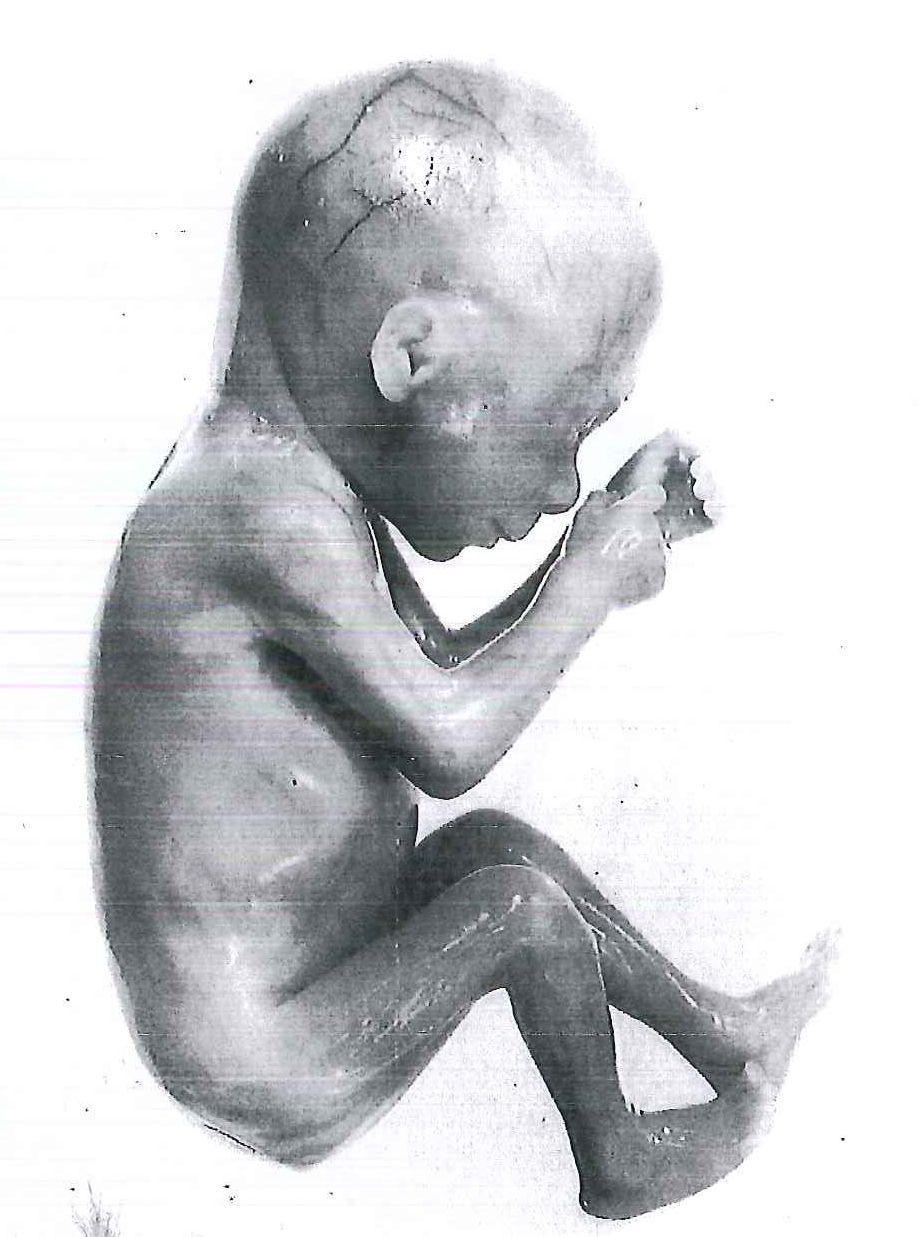
Figure 3. Photograph of a 17-week fetus. Note that the ears stand out from the head and that no hair is visible. Since there is no subcutaneous fat, the skin is thin, and the blood vessels of the head are visible. Fetuses at this age are unable to survive if born prematurely, mainly because their respiratory system is immature. The alveolar area is insufficient, and the vascularity of the lungs is underdeveloped.
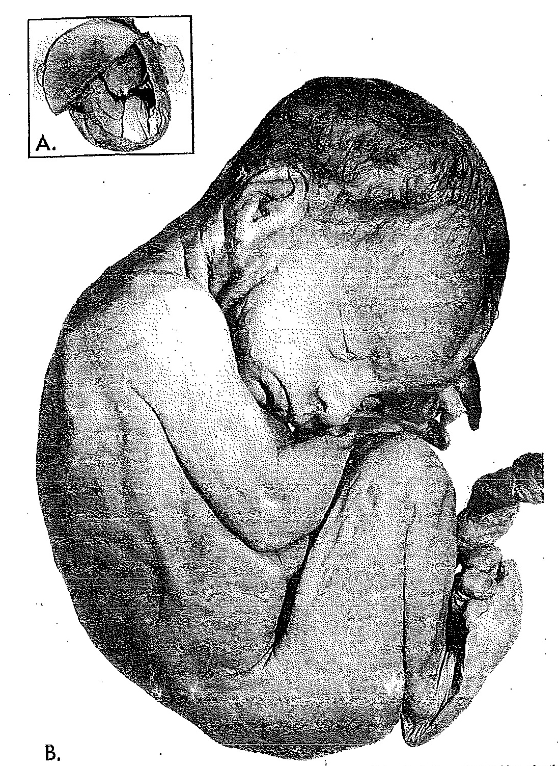
Figure 4. Photographs of a 25-week fetus. Note the wrinkled skin and rather lean body, resulting from the scarcity of subcutaneous fat. Observe that the eyes are beginning to open. A fetus of this size might survive if born prematurely, although it is not usually considered a viable fetus mainly because the lungs have not developed enough to provide adequate gas exchange.

